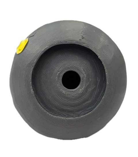
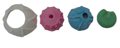
Below the plaster model on the middle shelf, we have 3D printed Contemporary Anatomy Teaching Models 1 and 2 and an accompanying braille sheet.
They were recently developed by Jonathan Shammas and Tammie Hunter, both Anatomy students, based on anatomical models from the Edinburgh University Anatomy Museum collection. They build on the technologies of their predecessors to make the anatomy of the eye more accessible through modern techniques.
Both models are replicas of the eyeball that can be disassembled like a Russian doll to explore 6 different internal structures of the eye. Here, the eyeball is enlarged to about the size of a tennis ball, and they are made from a durable, hard plastic so they can be handled as teaching aids.
Model 1 is displayed pieced together into a ball shape with the optic nerve protruding like a cable from its back. This model, along with the braille sheet, is displayed on the back half of the shelf, furthest away from us. In front of these is Model 2, which is displayed dissected into its six different parts.
Model 1 was developed first by Jonathan and was designed to be used in conjunction with an audio pen, with information that a user could listen to as they broke down the model. The information guides the user through each of the layers, their function and some common conditions that impact eyesight, which result from damage to these parts of the eye.
The goal of this model was to help blind and visually impaired people understand their conditions. However, Model 1 is very smooth textured, and all the layers were the same grey colour, making it difficult to feel the difference between them and limiting its utility.
Following on from this, Tammie developed Model 2, making some significant changes to the design. Here she moves away from the traditional colours employed by anatomy models like muted reds, creams and browns and instead opts for vibrant hues of grass green, sky blue, hot pink, sunshine yellow, coal black and bright white.
The 6 model parts are the same as Model 1, except that each layer is created with a distinct texture ranging from the delicate wavy grooves of the iris to the mountainous zigzag of bumps on the sclera. Instead of using an audio pen, Model 2 is accompanied by a Braille information sheet to explain more of the eye’s function.
As the models are significantly larger than life, closer to the size of a tennis ball than an eyeball, the Scottish Sensory Centre advised that, before exploring the models, it would be helpful to hold an object the same size as an eye to help with size perception. This led Tammie to use puffy sand a cost-effective material, which has a similar texture to an actual eyeball (though the real thing would be more jelly-like), to create a life-size eyeball for people to hold first, before using the scaled-up model.
These tactile models are an exciting innovation, and this process of adaptation could be used in any subject which relies on visual information for teaching. Such models can enrich learning experiences for everyone, not just students or people who are blind or visually impaired. |